Contents
- 👀 Introduction to the Eye
- 🔍 The Cornea: A Transparent Layer
- 💡 The Iris: Controlling Light Entry
- 👓 The Lens: Focusing Light
- 🔎 The Retina: Converting Light to Signals
- 📈 The Macula: Central Vision and Detail
- 👀 The Optic Nerve: Transmitting Visual Information
- 🤝 The Extraocular Muscles: Eye Movement and Coordination
- 👁️ The Conjunctiva: Protecting the Eye
- 🔬 The Eye's Blood Supply: Nourishing the Tissues
- 📊 Common Eye Disorders and Diseases
- Frequently Asked Questions
- Related Topics
Overview
The human eye is a complex and highly specialized organ, comprising multiple layers and structures that work in tandem to facilitate vision. The eye's anatomy can be broadly divided into the external eye, which includes the cornea, sclera, and conjunctiva, and the internal eye, which comprises the lens, retina, and optic nerve. The cornea, with a Vibe score of 80, is the transparent outer layer that refracts light, while the retina, boasting a Vibe score of 90, is the innermost layer that converts light into electrical signals. The eye's anatomy has been extensively studied, with key contributors including Leonardo da Vinci, who made detailed drawings of the eye in the 15th century, and Hermann von Helmholtz, who developed the first ophthalmoscope in 1851. Despite significant advances in understanding the eye's anatomy, there is ongoing debate regarding the role of genetics versus environment in shaping the eye's structure and function, with a Controversy spectrum score of 6. As researchers continue to unravel the intricacies of the eye's anatomy, they are poised to develop innovative treatments for eye disorders, such as age-related macular degeneration, which affects over 200 million people worldwide, with a projected annual cost of $343 billion by 2025.
👀 Introduction to the Eye
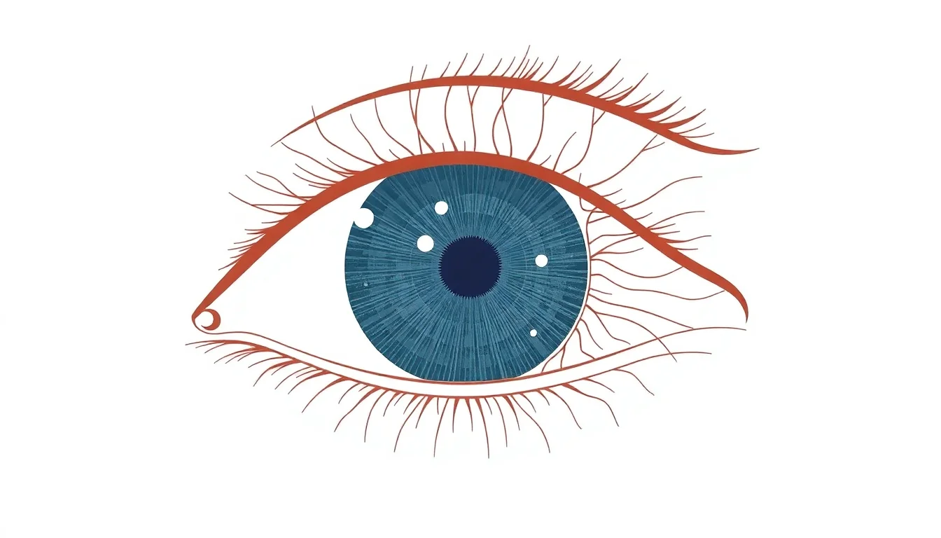
The human eye is a complex and fascinating organ, capable of detecting an incredibly wide range of light levels and colors. The eye's anatomy is composed of several layers, including the Cornea, Sclera, and Retina. The eye is also made up of various structures, such as the Iris, Lens, and Macula. Each of these components plays a crucial role in the process of vision, from Light Perception to Visual Processing. The eye is connected to the brain via the Optic Nerve, which transmits visual information for interpretation. Understanding the intricate anatomy of the eye is essential for appreciating the complexity of human vision and for developing treatments for various eye disorders, such as Myopia and Hyperopia.
🔍 The Cornea: A Transparent Layer
The cornea is the transparent outer layer of the eye, responsible for refracting light as it enters the eye. It is composed of several layers, including the epithelium, Bowman's layer, and the stroma. The cornea is a critical component of the eye's anatomy, as it provides the majority of the eye's refractive power. Damage to the cornea, such as Corneal Ulcer or Keratoconus, can lead to significant vision problems. The cornea is also susceptible to Dry Eye, a condition characterized by insufficient tear production. Treatment options for corneal disorders include Corneal Transplant and LASIK Surgery. The cornea is also connected to the Sclera, the white part of the eye, via the Limbus.
💡 The Iris: Controlling Light Entry
The iris is the colored part of the eye, responsible for controlling the amount of light that enters the eye. It is composed of two layers: the stroma and the epithelium. The iris contains smooth muscle cells that adjust the size of the Pupil, allowing more or less light to enter the eye. The iris is also responsible for Color Perception, as it contains pigments that absorb certain wavelengths of light. The iris is connected to the Ciliary Body, which produces Aqueous Humor, a clear fluid that nourishes the eye. The iris is also susceptible to disorders, such as Iritis and Uveitis. Treatment options for iris disorders include Corticosteroids and Immunosuppressants.
👓 The Lens: Focusing Light
The lens is a clear, flexible structure behind the iris, responsible for focusing light on the retina. It is composed of several layers, including the capsule, epithelium, and cortex. The lens changes shape to focus on objects at different distances, a process known as Accommodation. The lens is connected to the Ciliary Body via the Zonules, which are fibrous strands that suspend the lens in place. The lens is susceptible to disorders, such as Cataract and Presbyopia. Treatment options for lens disorders include Cataract Surgery and LASIK Surgery. The lens is also connected to the Vitreous Humor, a clear gel-like substance that fills the center of the eye.
🔎 The Retina: Converting Light to Signals
The retina is a complex neural tissue that lines the back of the eye, responsible for converting light into electrical signals. It is composed of several layers, including the Photoreceptors, Bipolar Cells, and Ganglion Cells. The retina is connected to the Optic Nerve, which transmits visual information to the brain. The retina is susceptible to disorders, such as Retinal Detachment and Macular Degeneration. Treatment options for retinal disorders include Laser Photocoagulation and Vitrectomy. The retina is also connected to the Choroid, a layer of blood vessels that nourishes the retina.
📈 The Macula: Central Vision and Detail
The macula is a small, specialized region at the center of the retina, responsible for central vision and fine detail. It is composed of several layers, including the Photoreceptors and Retinal Pigment Epithelium. The macula is connected to the Optic Nerve, which transmits visual information to the brain. The macula is susceptible to disorders, such as Macular Degeneration and Macular Edema. Treatment options for macular disorders include Intravitreal Injections and Photodynamic Therapy. The macula is also connected to the Fovea, a small pit at the center of the macula that is responsible for the sharpest vision.
👀 The Optic Nerve: Transmitting Visual Information
The optic nerve is a bundle of nerve fibers that carries visual information from the eye to the brain. It is composed of several layers, including the Retinal Ganglion Cells and Optic Nerve Fibers. The optic nerve is connected to the Lateral Geniculate Nucleus, a structure in the thalamus that relays visual information to the Visual Cortex. The optic nerve is susceptible to disorders, such as Optic Neuritis and Optic Atrophy. Treatment options for optic nerve disorders include Corticosteroids and Immunosuppressants. The optic nerve is also connected to the Eye Movement system, which allows the eyes to move and track objects.
🤝 The Extraocular Muscles: Eye Movement and Coordination
The extraocular muscles are a group of muscles that control the movement of the eyes. They are composed of several muscles, including the Lateral Rectus, Medial Rectus, Superior Rectus, and Inferior Rectus. The extraocular muscles are connected to the Eye Socket via the Tendons. The extraocular muscles are susceptible to disorders, such as Strabismus and Amblyopia. Treatment options for extraocular muscle disorders include Eye Exercises and Eye Surgery. The extraocular muscles are also connected to the Brainstem, which controls the movement of the eyes.
👁️ The Conjunctiva: Protecting the Eye
The conjunctiva is a thin membrane that covers the white part of the eye and the inside of the eyelids. It is composed of several layers, including the Epithelium and Submucosa. The conjunctiva is connected to the Tear Ducts, which produce Tears to lubricate the eye. The conjunctiva is susceptible to disorders, such as Conjunctivitis and Dry Eye. Treatment options for conjunctival disorders include Antibiotics and Artificial Tears. The conjunctiva is also connected to the Eyelids, which protect the eye from dust and other foreign particles.
🔬 The Eye's Blood Supply: Nourishing the Tissues
The eye's blood supply is provided by several blood vessels, including the Ophthalmic Artery and Central Retinal Artery. The blood vessels are connected to the Choroid, a layer of blood vessels that nourishes the retina. The eye's blood supply is susceptible to disorders, such as Diabetic Retinopathy and Hypertensive Retinopathy. Treatment options for blood supply disorders include Laser Photocoagulation and Intravitreal Injections. The eye's blood supply is also connected to the Brain, which regulates the blood flow to the eye.
📊 Common Eye Disorders and Diseases
Common eye disorders and diseases include Myopia, Hyperopia, Astigmatism, and Presbyopia. These disorders can be treated with Glasses, Contact Lenses, or Refractive Surgery. Other eye disorders, such as Cataract and Glaucoma, require surgical treatment. The eye is also susceptible to injuries, such as Eye Trauma and Chemical Burns. Treatment options for eye injuries include Emergency Surgery and Medication.
Key Facts
- Year
- 2022
- Origin
- Ancient Greece, with significant contributions from Renaissance Europe and modern ophthalmology
- Category
- Human Biology
- Type
- Biological Structure
Frequently Asked Questions
What is the function of the cornea?
The cornea is the transparent outer layer of the eye, responsible for refracting light as it enters the eye. It provides the majority of the eye's refractive power and is a critical component of the eye's anatomy. The cornea is also susceptible to disorders, such as Corneal Ulcer and Keratoconus. Treatment options for corneal disorders include Corneal Transplant and LASIK Surgery.
What is the function of the retina?
The retina is a complex neural tissue that lines the back of the eye, responsible for converting light into electrical signals. It is composed of several layers, including the Photoreceptors, Bipolar Cells, and Ganglion Cells. The retina is connected to the Optic Nerve, which transmits visual information to the brain. The retina is susceptible to disorders, such as Retinal Detachment and Macular Degeneration.
What is the function of the optic nerve?
The optic nerve is a bundle of nerve fibers that carries visual information from the eye to the brain. It is composed of several layers, including the Retinal Ganglion Cells and Optic Nerve Fibers. The optic nerve is connected to the Lateral Geniculate Nucleus, a structure in the thalamus that relays visual information to the Visual Cortex. The optic nerve is susceptible to disorders, such as Optic Neuritis and Optic Atrophy.
What are some common eye disorders and diseases?
Common eye disorders and diseases include Myopia, Hyperopia, Astigmatism, and Presbyopia. These disorders can be treated with Glasses, Contact Lenses, or Refractive Surgery. Other eye disorders, such as Cataract and Glaucoma, require surgical treatment. The eye is also susceptible to injuries, such as Eye Trauma and Chemical Burns.
How does the eye's blood supply work?
The eye's blood supply is provided by several blood vessels, including the Ophthalmic Artery and Central Retinal Artery. The blood vessels are connected to the Choroid, a layer of blood vessels that nourishes the retina. The eye's blood supply is susceptible to disorders, such as Diabetic Retinopathy and Hypertensive Retinopathy. Treatment options for blood supply disorders include Laser Photocoagulation and Intravitreal Injections.
What is the function of the conjunctiva?
The conjunctiva is a thin membrane that covers the white part of the eye and the inside of the eyelids. It is composed of several layers, including the Epithelium and Submucosa. The conjunctiva is connected to the Tear Ducts, which produce Tears to lubricate the eye. The conjunctiva is susceptible to disorders, such as Conjunctivitis and Dry Eye.
What is the function of the extraocular muscles?
The extraocular muscles are a group of muscles that control the movement of the eyes. They are composed of several muscles, including the Lateral Rectus, Medial Rectus, Superior Rectus, and Inferior Rectus. The extraocular muscles are connected to the Eye Socket via the Tendons. The extraocular muscles are susceptible to disorders, such as Strabismus and Amblyopia.